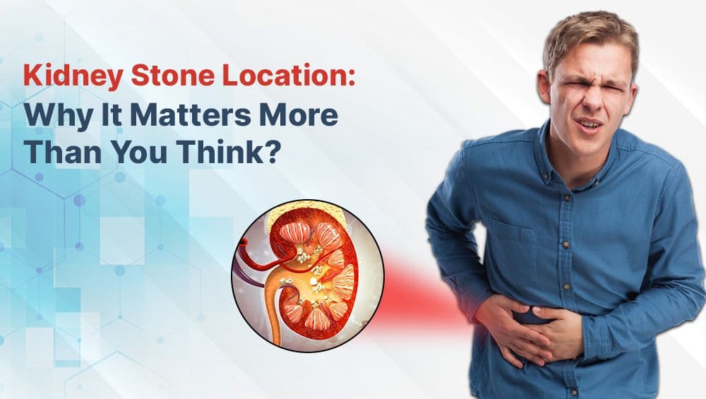
Kidney Stone Location: Why It Matters More Than You Think
Kidney stones occur when excess minerals and salts accumulate as hard deposits in the kidneys. Their size can vary from that of a grain of sand to a golf ball. While a lot is discussed regarding their causes, symptoms, and treatments, the location of the stone is one key issue that is often overlooked.
Kidney stones (renal calculi) develop when excess minerals and salts crystallize inside the urinary tract. While these hard mineral deposits can range in size from a grain of sand to a golf ball, their precise location within the urinary system plays a vital role in determining patient symptoms, diagnostic approaches, and surgical intervention strategies.
Whether a stone resides silently inside the kidney or migrates lower into the ureter or bladder, understanding its position is key to selecting the most effective treatment plan.
In this guide, we break down how kidney stone placement throughout the urinary tract affects pain levels, clinical evaluation, and advanced removal techniques.
The Urinary Tract: A Quick Overview
To understand why stone location matters, it helps to review the primary structures of the human urinary system:
- Kidneys: Two bean-shaped organs that filter waste and excess fluid from blood to produce urine.
- Ureters: Muscular tubes that transport urine from each kidney down to the bladder.
- Bladder: A hollow organ that stores urine until it is expelled.
- Urethra: The terminal channel through which urine exits the body.
Because the diameter of these passages narrows significantly from the kidney down to the urethra, even small stones can cause intense symptoms when migrating.
1. Stones in the Kidney (Renal Calyx and Renal Pelvis)
Kidney stones often form in the small cavities of the kidney (renal calyces) or the main collection area (renal pelvis). Non-obstructive stones resting inside the kidney often remain silent for long periods.
Typical Symptoms
- Mild, dull ache in the flank or back.
- Blood in the urine (hematuria).
- No noticeable symptoms if the stone is small and non-obstructive.
Diagnostic Evaluation
- Non-Contrast CT Scan: The gold standard for confirming stone position, density, and size.
- Renal Ultrasound: Highly effective for screening without radiation exposure.
- Abdominal X-Ray (KUB): Useful for tracking radiopaque calcium-based stones.
Treatment Approach
- Stones under 5 mm: Frequently pass spontaneously with conservative management and high fluid intake.
- Stones over 7 mm to 10 mm: Typically require intervention using flexible Retrograde Intrarenal Surgery (RIRS) or Extracorporeal Shock Wave Lithotripsy (ESWL).
- Large or Staghorn Stones (over 2 cm): Best managed via keyhole Percutaneous Nephrolithotomy (PCNL).
2. Stones in the Ureter (Ureteral Stones)
When a stone shifts from the kidney into the narrow ureter, it frequently becomes lodged at one of three anatomical transition zones:
- Ureteropelvic Junction (UPJ): Where the renal pelvis transitions into the ureter.
- Mid-Ureter: Where the ureter crosses over the pelvic brim.
- Ureterovesical Junction (UVJ): Where the ureter enters the urinary bladder.
Typical Symptoms
- Sharp, severe, wave-like pain radiating from the flank down to the lower abdomen and groin (renal colic).
- Persistent nausea and vomiting.
- Urgent, painful, or frequent urination.
- Pink, red, or brownish urine.
Diagnostic Evaluation
- Non-Contrast CT Scan: Provides immediate clarity on the exact blockage level and degree of kidney swelling (hydronephrosis).
- Targeted Ultrasound: Evaluates real-time urinary outflow obstruction.
Treatment Approach
- Stones under 5 mm: Managed conservatively with medical expulsive therapy (α-blockers), pain management, and heavy hydration.
- Stones over 5 mm or Obstructive Stones: Successfully treated with Ureteroscopy (URS) with Holmium/Thulium laser lithotripsy or flexible RIRS. Temporary DJ stent placement may be used to keep the ureter open during healing. Learn more about post-op management in our guide on how to avoid kidney stones recurrence.
3. Stones in the Bladder (Vesical Calculi)
Bladder stones are less common than kidney stones and generally form when urine stagnates in the bladder due to incomplete emptying—often triggered by an enlarged prostate (BPH), neurogenic bladder, or bladder diverticula.
Typical Symptoms
- Pain or discomfort in the lower abdomen (suprapubic area).
- Dysuria (painful urination) or terminal dribbling.
- Intermittent interruption of the urinary stream.
- Increased urge to urinate at night.
Diagnostic Evaluation
- Pelvic Ultrasound: Quickly visualizes stones floating or resting in the bladder base.
- Diagnostic Cystoscopy: Direct visual assessment using a flexible camera inserted through the urethra.
Treatment Approach
- Small Bladder Stones: May pass spontaneously with increased fluid consumption.
- Larger Bladder Stones: Removed using minimally invasive cystolitholapaxy, where endourological energy breaks the stone into fragments for vacuum extraction.
4. Stones in the Urethra (Urethral Stones)
Stones becoming trapped in the urethra represent a rare medical emergency. They typically occur when a migrating kidney or bladder stone becomes lodged on its way out.
Typical Symptoms
- Sudden, severe pain in the urethra or glans penis.
- Abrupt inability to pass urine (acute urinary retention).
- Visible redness or swelling around the urethral meatus.
Diagnostic Evaluation
- Clinical Examination: Physical assessment by an endourologist.
- Emergency Scanning: Rapid ultrasound or X-ray to confirm exact position.
Treatment Approach
- Endoscopic Retrieval: Precise dislodgement or laser fragmentation using delicate pediatric or adult endoscopes to clear the passage immediately.
How Stone Size and Location Interact
While location dictates symptom severity and surgical access, stone size determines whether procedural intervention is required:
- Stones smaller than 5 mm: High probability of spontaneous passage across all locations.
- Stones between 5 mm and 10 mm: Moderate chance of passage in lower locations, but frequently require URS or RIRS if stuck.
- Stones larger than 10 mm: Almost always require surgical intervention (RIRS, PCNL, or URS) to prevent severe kidney obstruction or long-term damage.
Preventive Steps for All Stone Locations
Regardless of where a stone forms or becomes lodged, core metabolic prevention strategies remain consistent:
- Hydration: Drink enough fluid to produce more than 2 liters of clear urine every day.
- Dietary Sodium Reduction: Keep daily sodium intake below 2,300 mg to reduce urinary calcium concentration.
- Balanced Animal Protein Intake: Moderate consumption of red meat and shellfish to lower uric acid levels.
- Pair Oxalates with Calcium: Combine dietary calcium (like dairy or yogurt) with high-oxalate foods during meals so oxalates bind in the digestive tract rather than the kidneys.
Consult Top Kidney Stone Specialist Dr. Deepanshu Gupta in Gurgaon
Accurate diagnosis of stone location is the most crucial step toward fast, painless relief and complete stone clearance.
Dr. Deepanshu Gupta at Cure Stone is a leading endourologist in Gurgaon and Delhi NCR with extensive clinical experience, having successfully performed over 9,500 endoscopic procedures. Specializing in advanced laser techniques—including radiation-free flexible RIRS, high-efficiency FANS RIRS suction laser surgery, URS, and Mini-PCNL—Dr. Gupta tailors every procedure to the exact position and density of your stone.
Don't let kidney stone pain compromise your health. Book a consultation with Cure Stone today for an accurate diagnosis and custom treatment plan!
Frequently Asked Questions (FAQs)
Why do ureteral stones cause more severe pain than kidney stones?
The ureter is a narrow, muscular tube with sensitive nerve networks. When a stone lodges in the ureter, it blocks urine outflow, causing pressure buildup in the kidney (hydronephrosis) and intense muscle spasms (renal colic).
Can a kidney stone be diagnosed with a simple ultrasound?
Ultrasound is an excellent, radiation-free tool for detecting stones in the kidney or bladder and checking for kidney swelling. However, a non-contrast CT scan is often necessary to pinpoint small stones lodged in the mid-ureter.
How do I know if a stone is about to pass?
As a stone approaches the lower ureter near the bladder (UVJ), symptoms often shift from severe back pain to intense bladder pressure, urinary frequency, and localized discomfort near the groin or urethra.
Have Questions About This Topic?
Get a personalized consultation at Cure Stone Hospital, Sector 52, Gurgaon.
Book Consultation