Hydronephrosis in USG Report: How to Understand Your Ultrasound Findings
Terms like "mild HUN with VUJ calculus" or "PCS dilated" can sound alarming or deceptively routine. This guide decodes a typical kidney ultrasound report line by line, so you know what each finding actually means.
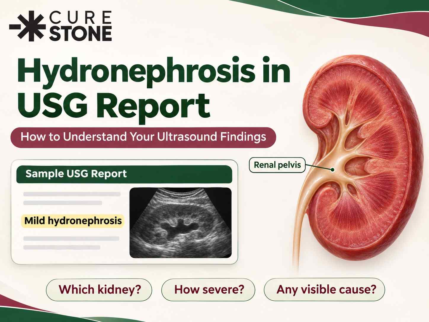
An ultrasound report is written for doctors, not patients. Phrases like "mild HUN with VUJ calculus", "PCS dilated" or "CMD maintained" can make a simple finding sound alarming, or make a serious one sound routine.
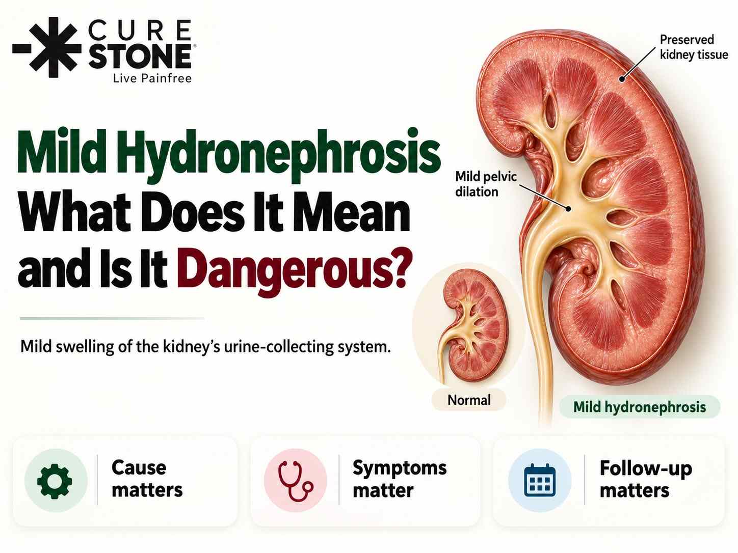
Short answer: When a USG report mentions hydronephrosis, it means the urine-collecting system inside the kidney looks swollen because urine is not draining freely. To understand what it means for you, read four things together: how much swelling (mild, moderate or severe), where the blockage seems to be, what is causing it (often a stone), and whether the kidney tissue itself looks healthy. The "Impression" at the end summarises this, but the lines above it tell you why.
This guide walks you through a typical kidney ultrasound report line by line, explains the common terms, and tells you what usually happens next for each type of finding.
What a Kidney (KUB) Ultrasound Looks At
A KUB ultrasound checks the Kidneys, Ureters and Bladder, and in men often the prostate. For each kidney, the radiologist usually reports:
- Size
- Thickness and brightness of the kidney tissue
- The urine-collecting system (where hydronephrosis is reported)
- Any stones, cysts or masses
- The ureter, if visible
- The bladder, and urine left behind after passing urine
Let's go through each.
Line by Line: Decoding Your Report
"Right kidney measures 10.6 x 4.8 cm"
Normal adult kidneys are roughly 9 to 13 cm long. A kidney that is clearly smaller than the other, or below the normal range, can suggest long-standing damage. A kidney that is larger than usual can be swollen from acute blockage or from other conditions. A difference of a centimetre or so between the two sides is common.
"Parenchymal / cortical thickness normal" or "cortex thinned out"
The parenchyma (cortex) is the working tissue of the kidney. Normal thickness is reassuring. "Thinned out cortex" or "cortical thinning" with hydronephrosis suggests the blockage has been present for a long time and has started to affect the kidney. This finding raises the urgency of treatment.
"Echogenicity normal" or "Raised echogenicity, Grade I / II / III"
Echogenicity is how bright the kidney tissue looks on the scan. Raised echogenicity is not hydronephrosis. It points towards "medical renal disease", meaning the kidney tissue itself is affected, for example by diabetes, high blood pressure or chronic kidney disease. It needs blood tests (creatinine, eGFR) and often a nephrologist's review. Read 10 signs of chronic kidney disease.
"Corticomedullary differentiation (CMD) maintained" or "CMD lost / poor"
A healthy kidney shows a clear difference between its outer layer (cortex) and inner layer (medulla). "Maintained" is normal. "Lost" or "poor" suggests the kidney tissue is diseased, which again calls for kidney function tests.
"PCS dilated" / "Mild hydronephrosis" / "Fullness of pelvis"
This is where hydronephrosis is reported. PCS means pelvicalyceal system, the collecting area inside the kidney. Radiologists commonly describe it like this:
| Report wording | What the radiologist sees | General meaning |
|---|---|---|
| Fullness of renal pelvis / prominent pelvis | Very slight widening, often borderline | Frequently not significant; may be a full bladder or anatomy |
| Mild hydronephrosis | Calyces enlarged but their shape preserved | Early or partial blockage |
| Moderate hydronephrosis | Calyces rounded, blunted | More significant blockage, needs evaluation |
| Severe or gross hydronephrosis | Ballooned calyces, very dilated pelvis, thin cortex | Severe or long-standing blockage |
Some reports also give a number for the renal pelvis AP diameter (in mm). A bigger number means more dilatation, but your doctor interprets it together with your symptoms, kidney function and the cause, not on its own.
"A 7 mm calculus seen in the lower calyx"
A calculus is a stone. The report gives its size and position:
- Upper, middle or lower calyx: inside the kidney, in its upper, middle or lower part
- Renal pelvis: in the central funnel of the kidney
- PUJ (pelviureteric junction): where the kidney joins the ureter, a common place for stones to get stuck
- Upper, mid or lower ureter: in the tube between kidney and bladder
- VUJ (vesicoureteric junction): where the ureter enters the bladder, the narrowest point and the most common place for a stone to lodge
Words such as "posterior acoustic shadowing" or "twinkling artefact" are features that support the diagnosis of a stone. Why the position matters so much is explained in kidney stone location: why it matters.
"Hydroureteronephrosis (HUN) up to VUJ" or "Proximal ureter dilated"
When the ureter is dilated too, the blockage is below the kidney. "Up to VUJ" means the ureter is swollen all the way down to the bladder, so the blockage is at or near the bladder entry, often a stone there. "Proximal ureter dilated" with a normal lower ureter points to a blockage higher up.
"Ureter not visualised"
This is common and usually not a problem. A normal, non-dilated ureter is thin and is often hidden behind bowel gas. It does not mean the ureter is missing or blocked.
"Urinary bladder: well distended, wall normal" / "Post-void residual (PVR): 20 ml"
The PVR is the urine left in the bladder after you pass urine. A small amount is normal. A large PVR, often with a thickened or trabeculated bladder wall and an enlarged prostate, suggests the bladder is not emptying well. This can cause hydronephrosis in both kidneys from back pressure. Read how bladder stones are removed if a stone is seen in the bladder.
"Back pressure changes"
This usually means swelling of both kidneys (and sometimes both ureters) from a problem at or below the bladder, such as an enlarged prostate, a narrowed urethra or a bladder that does not empty well.
"Bilateral"
Both sides are affected. Bilateral hydronephrosis, or hydronephrosis in a person with only one kidney, needs prompt evaluation because overall kidney function is at risk.
Other findings you may see
- Simple renal cyst: a fluid-filled bubble, very common with age and usually harmless. Read kidney cysts: symptoms and treatment.
- Parapelvic cyst: a cyst near the renal pelvis that can look like hydronephrosis on ultrasound.
- Extrarenal pelvis: a normal variation where the pelvis sits partly outside the kidney and looks roomy, without any blockage.
- Multiple cysts in both kidneys: may suggest polycystic kidney disease. Read polycystic kidney disease.
Reading the "Impression" Section
The impression is the radiologist's summary. Some common examples and what they generally mean:
| Impression | Plain meaning |
|---|---|
| "Right mild HUN with 5 mm right VUJ calculus" | A small stone is stuck where the right ureter enters the bladder, causing mild swelling above it |
| "Left renal calculus 8 mm, no hydronephrosis" | A stone sits in the left kidney but is not blocking urine flow at the moment |
| "Right moderate hydronephrosis, ureter not dilated, ? PUJ obstruction" | Blockage at the kidney-ureter junction is suspected; further tests needed |
| "Bilateral mild hydronephrosis with significant PVR and enlarged prostate" | Bladder not emptying well, causing back pressure on both kidneys |
| "Raised renal echogenicity, Grade II, suggestive of medical renal disease" | Kidney tissue is affected; not a blockage problem; kidney function tests needed |
| "Advised clinical correlation / CT KUB" | The ultrasound alone is not enough to decide; a CT or tests are recommended |
What Ultrasound Can and Cannot Tell You
Ultrasound is safe, quick and radiation-free, which is why it is usually the first test. But it has limits:
- It may miss small stones (under about 3 mm) and is not reliable for stones in the ureter, which bowel gas often hides.
- Stone size on ultrasound can differ from the size on CT.
- In the first hours of a blockage, especially if you are dehydrated, the kidney may not yet look swollen.
- A very full bladder during the scan can make the kidneys look mildly swollen; a repeat scan after passing urine may look normal.
- Cysts and an extrarenal pelvis can mimic hydronephrosis.
That is why a non-contrast CT KUB is often advised when you have pain, a stone is suspected but not seen, or treatment is being planned. It shows the exact size, position and density of the stone. Read the role of ultrasound and CT scan in diagnosing kidney stones and what to expect during your kidney stone tests.
What Usually Happens Next
| Your report shows | Usual next step |
|---|---|
| Mild fullness, no stone, no symptoms | Repeat scan after emptying the bladder, or follow-up ultrasound |
| Small stone (around 5 mm) in the lower ureter, mild hydronephrosis, pain controlled, no fever | Fluids, pain relief, sometimes medicine to relax the ureter, and a repeat scan |
| Stone 10 mm or more, or a stone that is not passing | Removal with URSL for ureteric stones or RIRS for kidney stones |
| Moderate or severe hydronephrosis, or cortical thinning | Urologist review soon, CT and kidney function tests, and often a renogram (DTPA or MAG3) to measure each kidney's function |
| Suspected PUJ obstruction | Renogram, then surgery if obstruction is confirmed |
| Bilateral hydronephrosis with high PVR | Urgent review of bladder emptying and kidney function |
| Raised echogenicity, CMD lost | Kidney function tests and nephrology review |
On the small stone row, read how long does it take to pass kidney stones. On the larger stone row, read about URSL, RIRS and best treatment for 10mm kidney stones. On suspected PUJ obstruction, read PUJ obstruction.
For the full picture of causes and treatment, read hydronephrosis causes and treatment and how to treat kidney blockage after stones.
When the Report Plus Your Symptoms Means Urgent Care
Whatever your report says, go to hospital the same day if you have hydronephrosis and:
- Fever, chills or shivering
- Pain not controlled by medicine
- Very little or no urine
- Persistent vomiting
- Only one kidney, or both kidneys affected
- Pregnancy with pain or fever
A blocked kidney with infection is an emergency regardless of the grade on the report. Read what is kidney infection (pyelonephritis) and can a stone in the ureter damage the kidney.
What to Bring to Your Consultation
- The full USG report and, if possible, the images or CD
- Any older scans, so changes can be compared
- Recent urine test, urine culture and creatinine results
- A list of your symptoms: where the pain is, fever, urine changes
- Your medicines, and whether you have diabetes, blood pressure, a single kidney or are pregnant
You can use our kidney stone checker to make sense of your stone size and position, explore kidney stone treatment options, or book a consultation with Dr. Deepanshu Gupta and bring your report for a clear explanation.
Frequently Asked Questions
What does "PCS dilated" mean in an ultrasound report?
PCS stands for pelvicalyceal system, the urine-collecting area inside the kidney. "PCS dilated" means this area is swollen, which is the same as hydronephrosis. The report usually adds whether it is mild, moderate or severe.
What does HUN mean in a USG report?
HUN means hydroureteronephrosis: both the kidney's collecting system and the ureter are swollen. It suggests the blockage is lower down, often near the bladder, for example a stone at the VUJ.
What is VUJ calculus?
It is a stone stuck at the vesicoureteric junction, where the ureter enters the bladder. This is the narrowest point of the ureter and the most common place for a stone to get stuck. Many small VUJ stones pass on their own; larger ones may need URSL.
Is "fullness of renal pelvis" the same as hydronephrosis?
It describes very slight widening, often borderline. It can be caused by a full bladder, extra fluid or normal anatomy. Your doctor will judge it with your symptoms and may repeat the scan after you pass urine.
Can ultrasound miss a kidney stone?
Yes. Ultrasound can miss small stones and is not reliable for stones in the ureter, which are often hidden by bowel gas. If symptoms suggest a stone and the ultrasound is unclear, a CT KUB is usually advised.
Does raised echogenicity mean hydronephrosis?
No. Raised echogenicity means the kidney tissue looks brighter than normal, which suggests the tissue itself is affected, for example by diabetes, blood pressure or chronic kidney disease. It needs kidney function tests, not stone treatment.
What does "ureter not visualised" mean?
It usually means the ureter is normal and not dilated, so it is too thin to see behind bowel gas. It is a common and generally reassuring phrase.
Should I worry if my report says mild hydronephrosis but I have no pain?
Not necessarily, but do not ignore it. Mild hydronephrosis without pain may be due to a full bladder, pregnancy or a slowly developing blockage. Show the report to a urologist, check your kidney function, and get a follow-up scan as advised.
References
1. Patti L, Leslie SW. Acute Renal Colic. StatPearls. Treasure Island (FL): StatPearls Publishing; updated December 2024. https://www.ncbi.nlm.nih.gov/books/NBK431091/
2. NYSORA. Hydronephrosis (Point-of-Care Ultrasound). https://nysora.com/pocus/5-1-hydronephrosis/
Have Questions About This Topic?
Get a personalized consultation at Cure Stone Hospital, Sector 52, Gurgaon.
Book Consultation