Non-Obstructing Kidney Stone on Ultrasound: Does It Need Treatment?
A non-obstructing kidney stone is not blocking drainage now. Learn when monitoring is reasonable, what follow-up means, and when treatment may be needed.

Your ultrasound report says “non-obstructing calculus in the kidney.” You may feel fine, yet the word “stone” raises an immediate question: does it need to come out now?
Short answer: A non-obstructing kidney stone is in the kidney without currently blocking urine drainage. If it is small and causes no symptoms, active surveillance may be a reasonable plan. That means follow-up imaging and a discussion of stone prevention, not forgetting it exists. Treatment becomes more relevant if the stone grows, causes pain or infection, or your circumstances make an unexpected stone episode difficult to manage. The decision depends on the full scan, your health and your preferences.
This guide explains what the scan wording means and how a urologist decides between monitoring and treatment.
What does “non-obstructing” mean?
A kidney has collecting spaces called calyces. A stone may sit in one of them while urine continues to drain through the kidney into the ureter. The report may describe it as a “non-obstructing renal calculus” or “calyceal stone.” It does not mean the stone can never move, grow or cause symptoms.
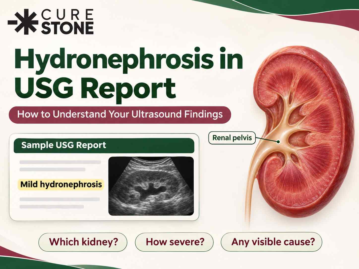
It also differs from a stone lodged in the ureter. A ureteric stone may block drainage and cause hydronephrosis, severe colic or infection. Check the report for words such as “dilatation,” “hydroureter” or “hydronephrosis,” and ask your doctor to interpret them in context. Our ultrasound report guide decodes several common terms.
Does a non-obstructing stone need surgery?
Not automatically. The American Urological Association says clinicians may offer active surveillance for an asymptomatic, non-obstructing calyceal stone, with follow-up imaging to look for growth or new stones. The European Association of Urology also describes observation as an option, while noting that the natural history is uncertain. These recommendations do not turn every small stone into a “safe forever” finding.
| Situation | What it may mean for the plan |
|---|---|
| Small stone, no pain, no infection or obstruction | Discuss surveillance and a follow-up scan. |
| Stone growing on successive scans | Review the interval and whether treatment is now worthwhile. |
| Recurrent pain or bleeding | Assess whether the stone is the cause; treatment may be considered. |
| Recurrent infection or a high-risk stone type | A more active approach may be needed. |
| One functioning kidney, poor access to care, or a job where sudden colic is hazardous | Discuss the risk of waiting more carefully. |
The right choice is individual. Stone size matters, but so do its position, growth, symptoms and your ability to attend follow-up.
Can it cause pain even without blockage?
Sometimes a patient has pain and a scan shows a non-obstructing stone, but the relationship is not always clear. A back or flank ache can also come from muscles, the spine or other organs. The AUA guideline notes that whether a non-obstructing calyceal stone causes pain is debated; treatment can be offered in selected cases after considering other causes, with no guarantee that removing the stone will remove the pain.
Tell your urologist when and where the pain occurs, whether it comes in waves, and whether you have urinary symptoms or fever. Other causes of kidney-stone-like pain are worth considering before choosing a procedure.
What does active surveillance involve?
It means agreeing on a follow-up plan, usually with imaging such as ultrasound or an X-ray when suitable. The interval is set by your clinician according to stone size, location, previous growth and your risk factors; there is no single schedule for everyone. CT may be useful when symptoms change or treatment is being planned, but repeated scans should balance information against radiation exposure.
Keep copies of the original and follow-up reports so the same stone can be compared over time. If you have had repeated stones, your doctor may discuss stone analysis and a 24-hour urine test to guide prevention. Drinking enough fluid for your circumstances and following personalised dietary advice are part of a prevention plan, but they do not reliably make a typical calcium stone disappear.
When might treatment be discussed?
Treatment can be reasonable if the stone grows, becomes symptomatic, is linked to recurrent infection, or waiting carries a practical risk for you. Options may include shock-wave lithotripsy or ureteroscopy/RIRS, depending on size, position, stone type and anatomy. RIRS versus other stone treatments explains how these approaches differ. A lower-pole stone has its own clearance considerations; see whether lower-pole stones pass naturally.
Do not schedule a procedure from the ultrasound phrase alone. A consultation should explain the expected benefit, the risks of intervention and the risks of waiting.
When should you seek urgent care?
New severe flank pain, fever or chills, vomiting that prevents you from drinking, very little urine, or feeling very unwell needs prompt assessment. A stone can move, and infection with obstruction is an emergency. If you have one functioning kidney or known kidney disease, seek advice early when symptoms change.
If you have a report but no clear plan, book a stone review and bring the images as well as the written findings.
Frequently Asked Questions
Is a non-obstructing stone harmless?
It may be quiet now, but it can grow, move or cause symptoms later. Ask for a monitoring plan rather than assuming it will always remain harmless.
Can a non-obstructing kidney stone pass on its own?
It may move out of the kidney and pass, particularly if small, but many stones remain in place. The chance depends on size and location and cannot be promised from one scan.
Does “no hydronephrosis” mean no treatment is needed?
No hydronephrosis is reassuring about current drainage, but treatment decisions also consider symptoms, growth, infection and personal circumstances.
How often should I repeat the ultrasound?
Ask the urologist who reviewed your scan. The timing depends on the stone and your risk; there is no universal interval.
Can water dissolve the stone?
Hydration supports stone prevention and may help passage if a small stone moves into the ureter. It does not dissolve most existing calcium stones. Some stone types, such as uric acid, need different, clinician-directed treatment.
References
- European Association of Urology. EAU Guidelines on Urolithiasis. Conservative management and indications for treatment of renal stones.
- American Urological Association. Surgical Management of Stones Guideline. Active surveillance for asymptomatic non-obstructing calyceal stones.
Have Questions About This Topic?
Get a personalized consultation at Cure Stone Hospital, Sector 52, Gurgaon.
Book Consultation