Mild Hydronephrosis: What Does It Mean and Is It Dangerous?
"Mild hydronephrosis" on an ultrasound report can mean a harmless full bladder or pregnancy, or a stuck stone that needs urgent care. See what decides whether it is dangerous, and which symptoms mean act now.
"Mild hydronephrosis" is one of the most common phrases on an ultrasound report, and one of the most misunderstood. Some people panic and assume kidney damage. Others ignore it because it says "mild". Both reactions can be wrong.
Short answer: Mild hydronephrosis means the urine-collecting area inside the kidney is slightly swollen, because urine is draining more slowly than normal. On its own it is often not dangerous, and it sometimes appears for harmless reasons such as a very full bladder during the scan or pregnancy. What decides whether it matters is the cause, your symptoms, how well your kidneys are working, and whether it persists. Mild hydronephrosis with fever, a single kidney, both kidneys affected, or a stone stuck in the ureter needs prompt attention.
This guide explains what the term means, the common harmless and not-so-harmless causes, which warning signs turn "mild" into urgent, and what tests and follow-up you should expect.
What Mild Hydronephrosis Means
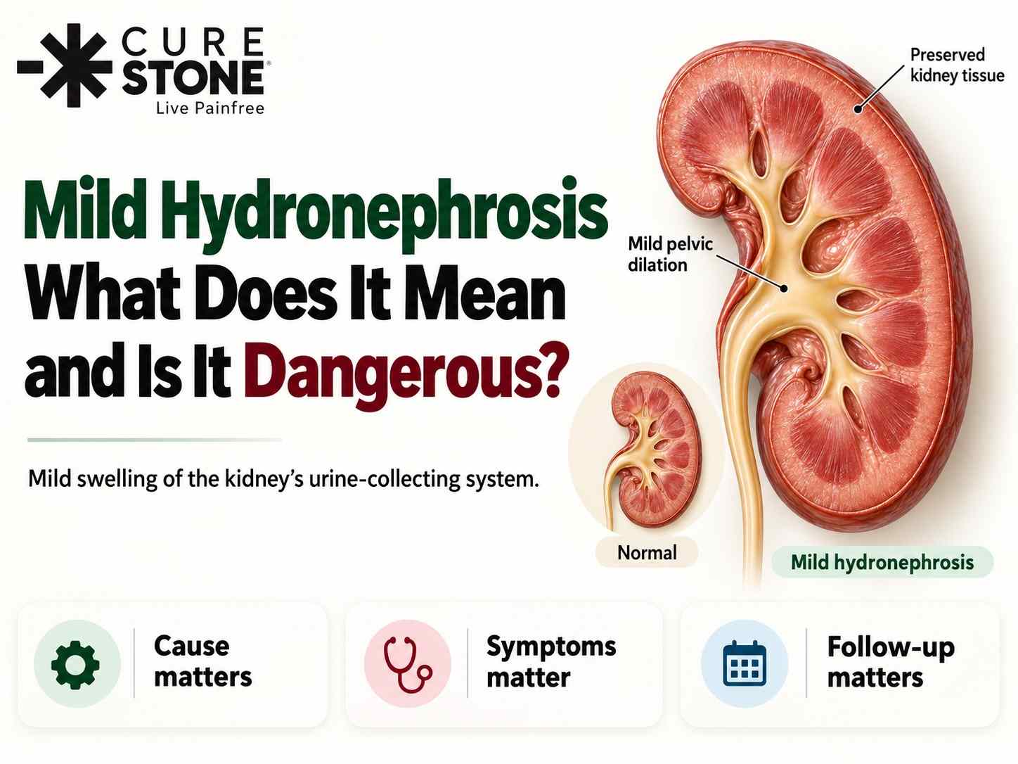
The kidney collects urine in small cup-shaped spaces called calyces, which drain into a central funnel called the renal pelvis, and then into the ureter towards the bladder. When urine flow is slowed or blocked anywhere along this path, urine backs up and these spaces stretch. That stretching is hydronephrosis.
Radiologists grade it by how much the collecting system is dilated:
| Grade on report | What the scan shows | What it usually means |
|---|---|---|
| Mild (often Grade 1 to 2) | Slight widening of the renal pelvis, calyces mostly normal shape | Early, partial or temporary slowing of drainage |
| Moderate (Grade 2 to 3) | Clear widening of the pelvis and calyces | More significant obstruction, needs evaluation |
| Severe or gross (Grade 4) | Marked swelling with thinning of the kidney tissue | Long-standing or severe blockage, risk of lasting damage |
Other terms you may see:
- PCS dilatation / pelvicalyceal dilatation: another way of describing hydronephrosis
- Hydroureteronephrosis (HUN): the ureter is swollen too, pointing to a blockage lower down, often near the bladder
- Fullness of the renal pelvis: very slight dilatation, often within normal limits
- Bilateral: both kidneys are affected
For the full overview of the condition, read hydronephrosis causes and treatment.
The Important Catch: "Mild" Does Not Always Mean "Minor"
The grade describes how swollen the kidney looks, not how serious the problem is. Two examples:
- A small stone that has just got stuck in the ureter may show only mild hydronephrosis in the first hours, especially if you are dehydrated, yet it can cause severe pain and, if infected, become an emergency.
- On the other hand, a long-standing mild dilatation that has not changed for years and causes no symptoms may need nothing more than an occasional check.
So the right question is not "how much swelling is there?" but "why is it there, and is the kidney working normally?"
Common Harmless or Temporary Reasons
Mild hydronephrosis sometimes shows up without any real blockage:
- A very full bladder during the scan. You are often asked to drink water and hold urine before an ultrasound. A tense bladder slows drainage from the kidneys and can make them look mildly swollen. A repeat scan after passing urine often looks normal.
- Pregnancy. Hormones relax the ureters and the growing uterus presses on them, more often on the right. In one study of pregnant women, 90% had at least mild dilatation on the right side. This usually settles after delivery. Read about kidney stones in pregnancy.
- High urine flow. Drinking large amounts of fluid just before the scan, or taking water tablets (diuretics), can temporarily distend the collecting system.
- Extrarenal pelvis. Some people are born with a renal pelvis that sits partly outside the kidney and looks roomy on a scan. It is a normal variation, not a blockage.
- A stone that has recently passed. The kidney can take a short while to return to normal after a stone comes out.
Causes That Need Attention
| Cause | Typical clues | Why it matters |
|---|---|---|
| Kidney stone in the ureter | Wave-like side or back pain, nausea, blood in urine | Can block the kidney and become infected |
| PUJ obstruction | Narrowing where the kidney joins the ureter, sometimes pain after drinking a lot | May slowly reduce kidney function |
| Enlarged prostate or urine retention | Older men, weak stream, incomplete emptying, often both kidneys affected | Back pressure can damage both kidneys over time |
| Ureteric stricture | Previous surgery, stone, injury or infection | Narrowing that can worsen with time |
| Vesicoureteric reflux | Mostly children, repeated urine infections | Urine flows back up towards the kidney |
| Compression from a mass | Pelvic or abdominal swelling, other symptoms | Needs investigation of the cause |
In adults, a kidney stone is the most common reason for new hydronephrosis with pain. Read do kidney stones cause urine blockage, PUJ obstruction, and can a stone in the ureter damage the kidney.
When Is Mild Hydronephrosis Dangerous?
Treat it as urgent, same day, if you have mild hydronephrosis and any of these:
- Fever, chills or shivering. An infected, blocked kidney can turn into sepsis within hours, whatever the grade. Guidelines recommend urgent drainage with a stent or a nephrostomy tube plus antibiotics, and delaying stone removal until the infection settles.
- Only one working kidney, or a kidney transplant
- Both kidneys affected (bilateral), especially with rising creatinine
- Very little or no urine
- Severe pain not controlled by medicine
- Pregnancy with pain, fever or urinary symptoms
Book a specialist review soon (within days) if:
- You have side or back pain that comes and goes
- There is blood in your urine
- A stone has been seen on the scan
- Your creatinine or other kidney blood tests are abnormal
- The swelling is new compared with an older scan
It is usually reasonable to monitor with a repeat scan if you have no symptoms, normal kidney function, two healthy kidneys, and a likely harmless reason such as a full bladder or pregnancy.
Read what is kidney infection (pyelonephritis) and UTI vs kidney infection.
What Tests Come Next?
Your doctor will choose tests based on your symptoms and history. The usual steps are:
- Repeat ultrasound after emptying the bladder, to rule out a full-bladder effect.
- Urine test and urine culture, to check for blood and infection.
- Blood tests (creatinine, urea), to check kidney function.
- Non-contrast CT KUB, if you have pain or a stone is suspected. It shows the exact size and position of any stone.
- Renogram (DTPA or MAG3 scan), if the dilatation persists without an obvious cause. It shows how much each kidney contributes to total function and whether urine is truly blocked or just draining slowly through a roomy system.
Read the role of ultrasound and CT scan in diagnosing kidney stones.
How Mild Hydronephrosis Is Treated
Mild hydronephrosis itself is not treated. The cause is treated, and the swelling settles once drainage improves.
- No clear blockage, no symptoms: observation and a repeat ultrasound, as your doctor advises.
- Small stone in the ureter, pain controlled, no infection: fluids, pain relief and sometimes medicine to relax the ureter, with a repeat scan to confirm it has passed. Read how long does it take to pass kidney stones and non-surgical treatment options for small kidney stones.
- Stone that does not pass, or is too large: removal with URSL for ureteric stones or RIRS for kidney stones, both through the natural urinary passage with no cut.
- Infection with blockage: urgent drainage with a DJ stent or nephrostomy and antibiotics first, stone treatment later. Read how to treat kidney blockage after stones.
- PUJ obstruction: surgery (pyeloplasty) if the renogram shows true obstruction or falling function.
- Prostate-related: medicines or prostate surgery to improve bladder emptying.
A stuck stone should not be "watched" indefinitely. Read what happens if you don't remove kidney stones and can I delay kidney stone removal.
Can Mild Hydronephrosis Go Away on Its Own?
Often, yes, when the cause is temporary: a full bladder, pregnancy, extra fluid, or a small stone that passes. Once drainage returns to normal, the kidney usually returns to its normal appearance.
If the cause persists, the swelling persists too, and a long-standing blockage can gradually reduce kidney function even without pain. That is why a repeat scan matters. Read can kidney stones cause permanent kidney damage.
Mild Hydronephrosis in Babies
Hydronephrosis is one of the most common findings on pregnancy scans, seen in roughly 1 to 5% of pregnancies. Most cases are mild, and many settle without treatment. In one follow-up study, about half of babies with mild antenatal hydronephrosis had a normal scan by 3 months. Babies diagnosed before birth should still have a follow-up ultrasound after birth, and some need checks for reflux or PUJ obstruction under a paediatric urologist or nephrologist.
Questions to Ask Your Doctor
- What is the most likely cause of my hydronephrosis?
- Is my kidney function normal?
- Is there a stone, and is it likely to pass?
- Do I need a CT scan or a renogram?
- When should I repeat the ultrasound?
- Which symptoms should make me come back urgently?
Get Your Report Reviewed
A report that says "mild hydronephrosis" deserves a clear answer, not guesswork. Use our kidney stone checker to understand your scan, explore kidney stone treatment options, or book a consultation with Dr. Deepanshu Gupta and bring your ultrasound or CT report. If you have fever with back pain, go to the nearest emergency department first.
Frequently Asked Questions
Is mild hydronephrosis serious?
Usually not by itself, but it depends on the cause. Mild hydronephrosis from a full bladder or pregnancy is often harmless. Mild hydronephrosis from a stuck stone, with fever, or in a single kidney needs prompt treatment.
Can mild hydronephrosis cause pain?
It can. Sudden blockage, for example from a stone, causes wave-like pain in the side or back that may spread to the lower abdomen or groin. Slowly developing mild hydronephrosis often causes no pain at all.
Can a full bladder cause mild hydronephrosis on ultrasound?
Yes. A very full bladder can temporarily slow drainage from the kidneys and make them look mildly swollen. A repeat scan after passing urine often clears up the question.
Can mild hydronephrosis lead to kidney failure?
Rarely, and only if a real blockage is left untreated for a long time, or if both kidneys or a single kidney are affected. With timely diagnosis and treatment of the cause, kidney function is usually preserved.
What should I eat or drink with mild hydronephrosis?
There is no special diet for hydronephrosis itself. Drink normally rather than forcing large amounts, especially if a stone may be blocking the ureter. If stones are the cause, a stone-prevention diet helps once the blockage is treated.
How often should mild hydronephrosis be rechecked?
It depends on the cause and your symptoms. Your doctor will usually repeat the ultrasound after a few weeks or months, or sooner if symptoms develop. A stone that has not passed needs earlier review.
Is mild hydronephrosis normal in pregnancy?
Mild dilatation, especially of the right kidney, is very common in pregnancy and usually settles after delivery. Pain, fever or urinary symptoms in pregnancy still need assessment for stones or infection.
Does mild hydronephrosis always need surgery?
No. Many cases need only observation or treatment of a temporary cause. Surgery is needed only when there is a blockage that will not resolve on its own, such as a stuck stone, a significant PUJ obstruction or a stricture.
References
1. Au KKL, Woo JSK, Tang LCH, Liang ST. Aetiological factors in the genesis of pregnancy hydronephrosis. Australian and New Zealand Journal of Obstetrics and Gynaecology. 1985;25(4):248-251. https://hub.hku.hk/handle/10722/184175
2. European Association of Urology. EAU Guidelines on Urolithiasis: Recommendations. https://uroweb.org/guidelines/urolithiasis/chapter/guidelines
3. D'Andrea V, Calabrese V, Ausili E, et al. Postnatal Follow-Up of Antenatal Hydronephrosis. Pediatric Research. 2010;68(Suppl 1):493 (conference abstract). https://www.nature.com/articles/pr20101199
Have Questions About This Topic?
Get a personalized consultation at Cure Stone Hospital, Sector 52, Gurgaon.
Book Consultation